Podiatry prides itself on measurement. We press-map, video-gate, laser-scan, and CAD-CAM like it’s second nature. Yet one of the biggest, most predictable sources of variance in the foot is hiding in plain sight: time. What your patient’s feet do at 8:00 am is not the same as what they do at 5:30 pm, because the entire neuromusculoskeletal and integumentary system is running on a 24-hour program. Understanding that daily rhythm—and designing your clinical decisions around it—can sharpen…
Author attribution not recorded
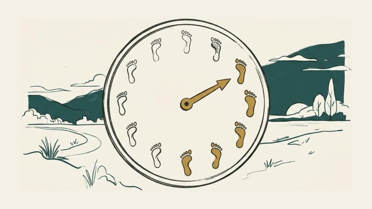
Podiatry prides itself on measurement. We press-map, video-gate, laser-scan, and CAD-CAM like it’s second nature. Yet one of the biggest, most predictable sources of variance in the foot is hiding in plain sight: time. What your patient’s feet do at 8:00 am is not the same as what they do at 5:30 pm, because the entire neuromusculoskeletal and integumentary system is running on a 24-hour program. Understanding that daily rhythm—and designing your clinical decisions around it—can sharpen diagnoses, stabilise your data, and improve outcomes. This is chronopodiatry in action. [1–3]
The daily physiology behind changing feet
Distal skin temperature follows a robust circadian rhythm, typically rising into the evening as peripheral vasodilation facilitates heat loss and sleep onset, and dipping in the biological morning. These cycles are clearly seen in continuous recordings from the hands and feet, and they differ with age and light exposure. [1,2] As thermoregulation and vascular tone fluctuate, hydrostatic forces during upright time drive progressive dependent fluid accumulation through the day. The result for most people—especially those who stand for work—is measurable diurnal swelling of the foot and ankle. Volumetry studies show morning-to-evening increases in otherwise healthy adults, and greater shifts in those with venous disease. [3–5] Those swelling curves co-travel with classic “heavy legs” and discomfort during prolonged standing. [5,6]
Sensory physiology also drifts across the day. Plantar cutaneous reflex pathways and radial measures of plantar sensitivity show time-of-day differences, and, more broadly, human pain sensitivity itself is under strong circadian control with a night-time peak and afternoon nadir. These effects are largely independent of sleep pressure and are big enough to matter clinically. [7,8]
Tendons—and their fibroblasts—keep time, too. Molecular clockwork within tendon acts as a peripheral oscillator regulating matrix homeostasis and collagen turnover; misalignment or damped rhythmicity is implicated in tendinopathy. Human tendon biopsies demonstrate rhythmic clock-gene expression, reinforcing that connective tissues are not “timeless” materials. [9,10] At the organ level, some work using shear-wave elastography in asymptomatic adults suggests Achilles tendon stiffness itself does not change meaningfully between morning and late afternoon when prior vigorous activity is controlled, but does transiently increase after a 30-minute run. That highlights behaviour (loading) as a key confounder atop any endogenous rhythm. [11]
Finally, postural control and gait parameters shift with time of day—and with sleep loss—according to syntheses across healthy and clinical cohorts. This is not only a laboratory curiosity; it changes fall risk windows, balance assessments, and context for interpreting “abnormal” performance. [12,13]
Do plantar pressures themselves change with time of day?
Direct head-to-head “8 am vs 6 pm” plantar-pressure studies are surprisingly scarce. What we do have is a large methodological footprint pointing to the same practical truth: plantar-pressure variables are highly sensitive to context. High-quality reliability papers typically collect on multiple days at the same time to minimise diurnal confounding, which is an implicit acknowledgement that time matters. [14,15]
We also know the foot’s geometry and stiffness are load-history dependent. In children, the Arch Height Index shows small but real intra-day variation and good test–retest reliability when measurement timing is standardised. [16] After sustained loading, such as distance running or long walking bouts, static arch height tends to be lower, and dynamic deformation greater, altering contact area and regional pressures even without any pathology. [17,18] Occupational cohorts with prolonged standing show end-of-day symptoms and measurable lower-limb volume gains that will redistribute loads inside a shoe. [6]
Put those strands together and a reasonable model emerges: what you measure is the sum of the person’s clock, their recent loading, and their fluid state. That’s why an afternoon scan after six hours on a retail floor looks and feels like a different foot to the pre-shift version—because it is. [3–6,16–18]
Why this matters for diabetic-foot risk and prevention
Diabetic foot disease hinges on repetitive, sub-perceptual mechanical trauma over time. The field is shifting from single snapshots of peak pressure toward “cumulative plantar tissue stress” (CPTS), integrating pressure, shear, and exposure time (steps, standing) and adherence to offloading. Daily rhythms and routines are therefore part of the aetiology: the after-lunch store walk-through, the evening cooking stint on tiles, the night-shift stocking round. Capturing dose across the waking day—rather than a single clinic pathway—tracks the true hazard. [19–22]
Shear matters as much as pressure for ulcer aetiology, and it also varies with footwear materials, temperature, gait velocity, and stance time—all of which can drift with time of day. Emerging reviews and experimental work continue to tie elevated shear and higher CPTS to ulceration and recurrence, while gentle daytime compression appears safe in selected diabetic patients with oedema and adequate arterial supply, reducing volume without compromising perfusion. [21,23–27]
The footwear and materials angle
If biological tissues have clocks, so do polymers—sort of. Midsole and insole foams (EVA, PU, TPU blends) are temperature-sensitive viscoelastic materials. Warmer environments reduce stiffness and increase damping; colder environments do the opposite. That means the same shoe or orthosis can feel and function differently across seasons and even across a single day as distal skin temperatures rise. Plantar soft tissues themselves are quasi-linear viscoelastic and rate-dependent, so the interface is a time- and temperature-dependent duet. [28–32]
Layer onto that the well-documented epidemic of poorly fitted footwear—and the simple fact that feet are larger in the evening due to diurnal swelling—and it becomes obvious why afternoon shoe fitting remains practical wisdom anchored in physiology. [3,30]
Chronopodiatry in clinic: practical implications
Standardise timing when you need comparability. If you are tracking change in plantar-pressure metrics, balance, or foot posture scores over time, schedule assessments at the same time of day and document recent loading (e.g., “no vigorous exercise 24 hours, <2,000 steps in prior 2 hours”). It’s exactly what many reliability studies already do. [14,15]
Decide whether you want the “fresh” foot or the “real-world” foot. For device provision, this is the pivotal choice. A morning capture might better represent an unloaded baseline and is often easier for casting or scanning tolerance. An afternoon capture may better reflect the foot as it lives—swollen after work, with lower arch height and different contact area—which could improve comfort and adherence in a worker on their feet. There is no universal rule here; the key is to choose deliberately and be consistent for follow-ups. [3,6,16–18]
Measure the day, not just the step. For people with diabetes at risk of ulceration, incorporate CPTS thinking. Use in-shoe sensors or wearable activity logs where possible, and counsel to spread standing tasks, sit when safe, and adhere to offloading especially at “high-dose” times of their day. [19–22]
Account for materials changing with temperature. When queueing lab testing or athlete fit-ups, keep test temperatures consistent and note recent cold/hot exposure. Educate patients that midsoles and insoles may feel stiffer on cold mornings and softer late in a hot day, and that this can nudge symptoms. [28,29,31,32]
Schedule rehabilitation with the clock. Pain sensitivity tends to peak overnight and is lower in the mid-afternoon. For some patients, that may influence when to start graded loading or when to attempt more provocative exercises, especially early in programs. Conversely, for inflammatory arthritides with pronounced morning stiffness, expect diurnal changes in gait velocity and comfort that may shape appointment timing and orthoses break-in. [8,33]
Balance testing and night-shift realities. Distal skin-temperature rhythms shift with light and schedule. Night-shift workers will bring a different physiology to your 9 am clinic than day-workers. Document shift status; if you are monitoring change, keep the same relative biological timing where feasible. [1,34]
Research gaps that podiatry can help close
We need well-powered within-subject studies directly comparing morning vs afternoon plantar-pressure fields in representative populations (standing workers, runners, people with diabetes), with concurrent limb volume, distal skin temperature, and activity control. We need mechanistic work linking diurnal oedema to shear hotspots in shoes and boots. We need trials testing whether time-standardised capture improves the signal-to-noise of orthoses optimisation. And we need pragmatic CPTS-based protocols that integrate “when” into “how much.” Until then, podiatry can still reap dividends by bringing time of day into our documentation, protocols, and conversations with patients.
Bottom line
Your pedobarograph doesn’t just measure a foot; it measures a foot at a time. The vascular, neural, connective, and material subsystems that shape plantar loading all run to a daily beat. Control the timing when you need precision, embrace the afternoon foot when you need real-world comfort, and integrate the day’s dose when you are protecting skin. Chronopodiatry won’t add minutes to your appointments, but it will add meaning to your numbers. [1–34]
References
Kantermann T, Ghotbi N, Schmid SM, et al. Circadian rhythm of distal skin temperature in healthy older and young women. Clocks & Sleep. 2024;6(4):102. doi:10.3390/clockssleep6040102. MDPI
Fernandes AA, Marins JCB. Daily variation of skin temperature in humans through infrared thermography: a pilot study. J Therm Biol. 2017;70:330-336. ScienceDirect
Ashizawa K, Kumai T, Kusumoto A, et al. Diurnal variations in volume of the foot and ankle. Foot Ankle Int. 2001;22(7):575-578. ScienceDirect
Rabe E, Pannier F, Ko A, et al. Venous insufficiency in male workers with a standing profession: leg volume at the beginning and end of workdays. Dermatology. 2009;219(2):121-127. Karger
Ziaja D, Simka M, et al. Venous leg symptoms in healthy subjects assessed during prolonged standing. J Vasc Surg Venous Lymphat Disord. 2017;5(1):25-32. JVS Venous
de Lima Cavalcante N, et al. Musculoskeletal disorders of the foot and ankle among nurses: a systematic review. SAGE Open Med. 2023;11:20503121231159520. SAGE Journals
Coroian A, et al. Diurnal changes in plantar sensitivity. Neurosci Lett. 2011;502(3):181-185. ScienceDirect
Daguet I, Raverot V, Bouhassira D, Gronfier C. Circadian rhythmicity of pain sensitivity in humans. Brain. 2022;145(9):3225-3235. doi:10.1093/brain/awac147. Oxford Academic
Ali O, Millar NL. Role of the tendon circadian clock in tendinopathy and implications for therapy. Int J Exp Pathol. 2024;105(3):101–111. Wiley Online Library
Boesen AP, Dideriksen K, Kjær M. Investigating circadian clock gene expression in human tendon biopsies. Eur J Appl Physiol. 2019;119(8):1735-1744. SpringerLink
Chortis N, Webborn N, et al. Shear wave elastography measures of the Achilles tendon: influence of time of day, leg dominance and a 30-minute run. Appl Sci. 2018;8(7):1170. MDPI
Horiuchi M, et al. The effect of time-of-day and sleep deprivation on postural control: a systematic review. Gait Posture. 2022;96:80-92. ScienceDirect
Arciero E, et al. Does time of day influence postural control and gait? A review. Gait Posture. 2021;90:30-38. ScienceDirect
Kernozek TW, et al. Between-day reliability of repeated plantar pressure distribution measurements. Gait Posture. 2007;26(2):197-201. Europe PMC
Sun D, Zhou X, et al. Normal foot loading parameters and repeatability of the Footscan system. J Foot Ankle Res. 2017;10:35. BioMed Central
Hillstrom HJ, et al. Reliability and convergent validity of Arch Height Index and Arch Rigidity Index in children; intraday differences at two time points. J Am Podiatr Med Assoc. 2017;107(5):—. japmaonline.org
Hamuro T, et al. Relationship between medial longitudinal arch and running form; post-run static arch height decreases. Biomechanisms. 2016;24:169-176. jstage.jst.go.jp
Wu W, et al. Running/walking distance and plantar pressure distribution across the day. BMC Musculoskelet Disord. 2022;23:—. BioMed Central
Bus SA, et al. New biomechanical models for cumulative plantar tissue stress. J Biomech. 2024;—. ScienceDirect
Waaijman R, et al. Measuring plantar tissue stress in people with diabetic peripheral neuropathy: a critical concept. J Diabetes Sci Technol. 2019;13(4):—. SAGE Journalsdiabetesfeetaustralia.org
Savage R, et al. Plantar shear stress in the diabetic foot: systematic review and meta-analysis. Diabet Med. 2020;37(4):—. Wiley Online Library
Rana S, et al. Plantar pressure and shear during gait in diabetic peripheral neuropathy. J Clin Transl Endocrinol. 2025;—. SpringerLink
Partsch H, et al. Safety and efficacy of mild compression (18–25 mm Hg) therapy in patients with diabetes and lower-extremity oedema. J Diabetes Sci Technol. 2012;6(3):641-647. SAGE Journals
Blazek C, et al. Oedema management in a diabetic patient with foot ulceration and PVD: case study. Diabet Foot J. 2016;19(1):38-42. DiabetesontheNet
Ulbrecht JS, et al. Risk factors for plantar ulcer recurrence in neuropathic diabetes. Diabetes Care. 2014;37(6):1697-1705. Diabetes Journals
Cheung JT-M, Zhang M, et al. Compressive material properties of plantar soft tissue. J Biomech. 2007;40(1):—. ScienceDirect
Ledoux WR, Meaney DF, Hillstrom HJ. A quasi-linear, viscoelastic structural model of plantar soft tissue. J Biomech Eng. 2004;126(6):831-837. seas.upenn.edu
Wang Y, et al. Effects of temperature on midsole and insole materials. Mater Des. 2011;32(10):—. ScienceDirect
Tsai C-J, et al. Mechanical characteristics of EVA foams with different compositions. Mater Sci Eng C. 2022;—. ScienceDirect
Hawke F, Burns J, Radford JA, du Toit V. Incorrectly fitted footwear, foot pain and disorders: systematic review. J Foot Ankle Res. 2018;11:43. BioMed Central
Kim J, et al. Temperature effects on impact and rebound properties of footwear midsoles. Korean J Mater Res. 2010;20(10):—. Earticle
Liao Y, Zhang X, et al. In-vivo dynamic mechanical analysis of plantar soft tissues. Technologies. 2025;13(5):191. MDPI
Carroll M, et al. Gait velocity exhibits >50% diurnal variation in rheumatoid arthritis. J Foot Ankle Res. 2010;3(Suppl 1):P1. BioMed Central
Krauchi K, et al. Skin-temperature rhythms respond to night-shift schedule and light. J Biol Rhythms. 2017;32(2):—. SAGE Journals
Notes: Where page ranges are not listed in the digital object or abstract landing pages, they are denoted “—” to avoid incorrect pagination.