If you see kids, you see heel pain. Football boots and netball courts are back, training loads spike, and suddenly half your afternoon clinic is limping through the door. Too often, the label is instantaneous—“Sever’s”—and the plan is a one-size-fits-all script of heel cups, stretches and a sabbatical from sport. Sometimes that’s right; often it’s lazy. This piece takes you past reflex labels and into a structured, evidence-based, humane approach you can use tomorrow. It’s written for…
Author attribution not recorded
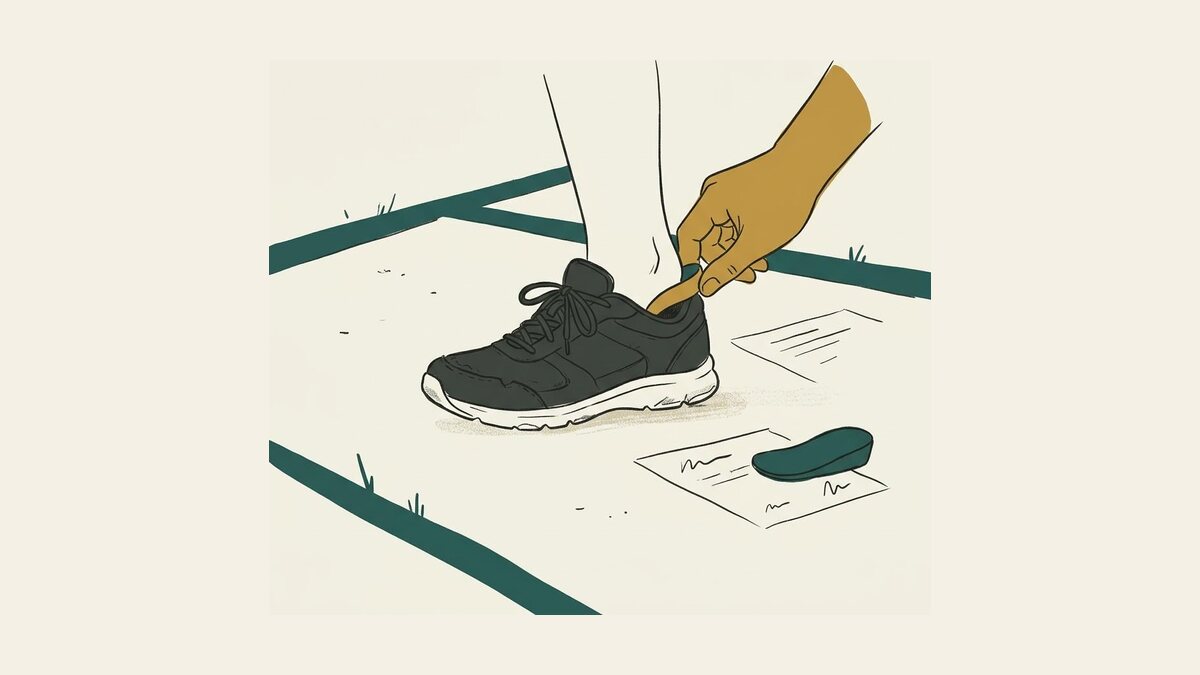
If you see kids, you see heel pain. Football boots and netball courts are back, training loads spike, and suddenly half your afternoon clinic is limping through the door. Too often, the label is instantaneous—“Sever’s”—and the plan is a one-size-fits-all script of heel cups, stretches and a sabbatical from sport. Sometimes that’s right; often it’s lazy. This piece takes you past reflex labels and into a structured, evidence-based, humane approach you can use tomorrow. It’s written for podiatrists and allied clinicians, but pitched so an engaged parent can follow along too.
A 60-second story to start
Luca is 10, left-foot dominant, plays soccer and basketball, and has just jumped two training squads. Three weeks into the new schedule he’s hobbling by half-time, then tip-toeing the morning after. Squeeze the heel and he winces. The instinct is “calcaneal apophysitis” (the growth plate is grumpy), reassure, modify load, add a heel lift and a stretch sheet, and wish him luck. That might work. But if he’s still sore in four weeks—or if there’s night pain, focal bony tenderness, or a rigid flatfoot—you’ll wish you’d cast a wider net earlier. The good news? A clear, stepwise approach catches what matters and keeps most kids moving safely.
What are we really talking about?
Calcaneal apophysitis (historically “Sever’s disease”) is irritation of the calcaneal growth plate, typically in active, peri-pubertal kids during spikes in running and jumping. It’s common, self-limited, and usually clinical—no imaging needed in straightforward cases. Risk factors cluster around sudden load increases, multi-sport participation, and (to a lesser extent) higher BMI; the literature continues to evolve on intrinsic and extrinsic contributors. (1,2)
But paediatric heel pain is a family of problems, not a single diagnosis. Your differential should always include: calcaneal bone stress injury, retrocalcaneal bursopathy, Achilles insertional tendinopathy (less common but not rare), tarsal coalition (classically later childhood with a rigid flatfoot), neural entrapment, infection, and very rarely tumour. The most important skill is triage: who is safe for conservative care now, and who demands a different path today. (3)
Red flags you must not miss
Night pain, systemic symptoms, or rest pain out of proportion to activity.
Acute inability to weight bear or fever—always think infection/septic arthritis in limping children.
Point bony tenderness over the calcaneus after a big load spike: treat as possible stress injury until proven otherwise.
Rigid flatfoot, recurrent ankle sprains or peroneal spasm around age 12–16: think tarsal coalition. (3–5)
Examination that buys you certainty
Observation and gait. Antalgic gait, toe-walking to unload the heel, and early-morning “first-step” behaviours are informative. (1)
The calcaneal squeeze test. Mediolateral compression of the posterior calcaneus typically reproduces apophyseal pain. Use it, but don’t let it blind you to deeper bony pain from stress injury. (6)
Load-provocation and cuff tests. Hopping and heel raises: pain patterns and capacity (reps, symmetry) help profile irritability and readiness to progress. (7)
Foot posture and mobility. Check rearfoot motion. Limited subtalar movement with a valgus, rigid flatfoot should trigger coalition thinking. (5)
When to image: in classic apophysitis, you don’t need it; X-rays often show “changes” that are common in pain-free kids. Image only to rule out other pathology (fracture, stress injury, coalition) or if the story is atypical, severe, or not improving as expected. (1,8)
What the best evidence actually says (and doesn’t)
A large factorial randomised trial (n≈124) compared heel raises vs prefabricated orthoses and “new supportive shoes” vs current shoes. At two months, heel lifts had a small short-term edge; by 6–12 months, outcomes converged—everyone improved, regardless of insert or footwear swap. In other words: kids generally get better; simple cushioning/elevation may help in the short term, but there is no long-term magic device. (9,10)
A 2017 evidence review for family physicians reached a similar conclusion: inserts may give modest short-term relief; by three months, most children improve with or without specific devices. (10)
More recent systematic reviews reiterate that conservative care is first-line and that research heterogeneity remains high—load management, symptom-led progression, and common-sense supports still anchor care. (2)
Quality-of-life research reminds us this hurts more than a little: calcaneal apophysitis measurably dents a child’s daily functioning, and parents often underestimate the impact. Don’t trivialise it. (11)
The practical plan (what you’ll say and do)
1) Name the problem and set expectations
Explain the growth-plate story in plain language, emphasising that the bone is temporarily vulnerable while the rest of the kinetic chain is trying to keep up with growth and load. Most cases settle with measured adjustments; many will recur with load spikes until the apophysis matures. That’s normal, not failure. (1)
2) Modulate load, don’t ban sport
The goal is pain-guided activity, not bubble-wrap. Use a simple traffic-light rule: green (0–2/10 pain, no next-day fallout), amber (3–5/10, modify drills/minutes), red (≥6/10, stop impact for now). Allow skills, passing drills, and positions with lower running demand while temporarily trimming sprints, jumps and back-to-back sessions. This preserves identity, conditioning, and compliance.
3) Cushion and (maybe) lift
A shock-absorbing heel cup or small heel lift often helps in the short term, especially in studs or minimal turf shoes. It lowers peak heel forces and reduces Achilles traction during the most irritable window. Don’t oversell brand or model—comfort, fit and practical use beat theory. Expect the benefit to normalise over months. (10)
4) Strength and capacity—not just “stretch your calves”
Tight calves matter, but long, sleepy calves matter more. In the quiet phase, start gentle gastrocnemius–soleus stretching and progressive heel raises (bilateral → single-leg; floor → step; straight-knee and bent-knee). Add hip and foot intrinsic work. Progressions should be criteria-based, not calendar-based: when a child can perform 25 pain-free single-leg heel raises and complete 30 minutes of brisk walking without next-day reset, you can begin impact re-introduction. (7)
5) Footwear reality check
Supportive, well-cushioned trainers for school and cross-training protect the heel between sport exposures. Shoe replacement alone didn’t outperform usual shoes in the RCT, so treat it as a comfort adjunct, not a cure. For boots with stiff soles and low pitch, a discreet heel cup often makes the biggest difference. (9)
6) Analgesia and short-term off-loading
Ice can help an irritable heel after sessions. If walking is painful, a CAM walker boot for one to two weeks provides relief while you reset load—especially in kids who cannot “not play” without a firm boundary. Reserve casts for rare, severe cases or proven bone stress; they’re seldom needed. (1)
7) A staged return to running and sport
Use a simple, graded running program over 3–5 months in higher-irritability cases, blending intervals, hops, then multi-plane drills before full training and games. Advance only if the child meets criteria and has no next-day flare. This is overkill for many but gold for the serially sore. (7)
When it’s not calcaneal apophysitis
Tarsal coalition
Think older child, insidious lateral hindfoot pain, recurrent sprains, limited subtalar motion, and a rigid valgus flatfoot that doesn’t “resupinate” on heel raise. Start with weight-bearing oblique radiographs; CT/MRI may be needed. Early care is activity modification, orthoses, and peroneal calm-down; persistent, function-limiting pain may warrant surgical resection in selected cases. Don’t miss it—and don’t mistake it for stubborn “Sever’s.” (5,12)
Calcaneal bone stress injury
Watch for night pain or focal bony tenderness with a load-spike story. X-rays can be normal early; MRI is the best test if suspicion is high. Management is load cessation/off-loading, then a structured re-build—rushing this is how you convert irritability into a season-ending fracture. (3)
Infection and tumour (rare, vital)
Fever, malaise, erythema, systemic symptoms, or persistent deep night pain demand urgent work-up. The child who “can’t walk today” with a hot, swollen joint is never a watch-and-wait situation. (4)
Talking to parents (so they become allies, not skeptics)
Validate the pain and the child’s distress. Quality-of-life data show meaningful impact—kids aren’t being dramatic. (11)
Explain why you’re not scanning “to be safe” in classic cases: imaging commonly shows normal variants that don’t change care. (1,8)
Frame the plan as a set of dials (load, cushion, capacity, footwear, time) you’ll turn up and down together.
Forecast the recurrence pattern around growth spurts and season transitions so nobody panics when it flickers back. (1)
A clinic-ready algorithm (in words, not boxes)
Screen for red flags and atypical features. If present, image/refer accordingly. If not, proceed. (4)
Diagnose clinically. Posterior heel pain in an active child, positive squeeze, load-linked symptoms: likely apophysitis. (6)
Start conservative care: load modulation, heel cup/lift, strength and stretch plan, footwear check, parent education. (9,10)
Reassess at 2–4 weeks. Improving: progress capacity and a graded return. Stalled/worse: reconsider differential; think coalition or stress injury. (5)
Escalate thoughtfully. CAM boot for short spells if walking is painful; imaging if features are atypical or progress stalls despite adherence. (1)
Myths worth retiring
“Stretching fixes Sever’s.” Stretching can help, but strength and total load are usually the bigger levers. (7)
“Get new shoes; problem solved.” Shoe swaps alone don’t outperform current shoes long-term. Comfort matters more than marketing. (9)
“If it still hurts in two weeks, we’ll get an X-ray.” Imaging rarely helps in classic apophysitis and can mislead. Save it for red flags or when the story doesn’t fit. (1)
Take-home for busy clinics
Paediatric heel pain isn’t a single diagnosis. Rule out the dangerous and the different, then treat the common well. (3)
Evidence supports simple, conservative care with short-term cushioning benefits and long-term convergence of outcomes. Manage load, build calf and hip capacity, and keep kids connected to sport. (9,10)
Use boots sparingly, casts rarely, and imaging judiciously. Most kids don’t need them; some absolutely do. Knowing which is your craft. (1)
References
American Academy of Orthopaedic Surgeons. Sever’s disease (heel pain). OrthoInfo. Last reviewed Jun 2022. OrthoInfo
Nieto-Gil P, Marco-Lledó J, García-Campos J, Ruiz-Muñoz M, Gijon-Nogueron G, Ramos-Petersen L. Risk factors and associated factors for calcaneal apophysitis (Sever’s disease): a systematic review. BMJ Open. 2023;13(6):e064903. BMJ Open
Rio E, Mayes S, Cook J. Heel pain: a practical approach. Aust Fam Physician. 2015;44(3):96-101. RACGP
Royal Children’s Hospital Melbourne. Clinical practice guideline: the limping or non-weight bearing child. Royal Children's Hospital
Orthobullets. Tarsal coalition – Pediatrics. Updated May 3, 2024. Orthobullets
Radiopaedia. Calcaneal apophysitis (reference article). Last revised 2025. Radiopaedia
Massachusetts General Brigham Sports PT. Pediatric rehabilitation protocol for calcaneal apophysitis (Sever’s disease). 2024. Massachusetts General Hospital
Volpon JB, de Carvalho Filho G. Calcaneal apophysitis: a quantitative radiographic evaluation of the secondary ossification center. Arch Orthop Trauma Surg. 2002;122(5):338-341. SpringerLink
James AM, Williams CM, Haines TP. Effectiveness of footwear and foot orthoses for calcaneal apophysitis: a 12-month factorial randomised trial. Br J Sports Med. 2016;50(20):1268-1275. British Journal of Sports Medicine
Uvelli K, Neher JO, Safranek S. Treatment for calcaneal apophysitis. Am Fam Physician. 2017;96(2):126-127. AAFP
James AM, Williams CM, Haines TP. Health-related quality of life of children with calcaneal apophysitis: child & parent perceptions. Health Qual Life Outcomes. 2016;14:95. BioMed Central
Kothari A, Masquijo J. Surgical treatment of tarsal coalitions in children and adolescents. EFORT Open Rev. 2020;5(2):80–89. doi:10.1302/2058-5241.5.180106. (Original publication). PubMed